
Peat as an Adsorbent for Water Purification in Developing Countries

FTIR Analysis

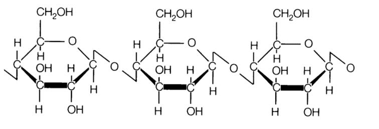
Figure 1: FTIR Spectrum of Sphagnum Moss Peat
Figure 2: Molecular Structure of Cellulose
Figure 1 shows the FTIR Spectrum of peat that has not been contacted with Pb2+ or Zn2+ ions. There are 3 main functional groups that were identified from the spectrum, namely the O-H stretch, C=C stretch as well as the C-O-C stretch. These functional groups can be observed in the structure of cellulose as shown in Figure 2.

Figure 3: Overlay FTIR Spectra of Peat and Peat contacted with Lead(II) ions
Figure 4: Comparison of wavelengths
Figure 3 shows the overlay of two FTIR spectrums. The red line shows the spectrum of peat whereas the blue line shows the spectrum of peat that has been contacted with Lead(II) ions. As indicated by the box, there was a shift in the wavelength of the O-H stretch from 3371cm-1 to 3403 cm-1. The wavelength of peat that has been contacted with Zn2+ also showed an increase in wavelength to 3407 cm-1. This shows that the hydroxyl group (O-H) is the main functional group that is responsible for the adsorption of the metal ions. The wavelength of the other two functional groups do not shows a major change in wavelength.
.
.